What Is Pericardial Effusion
Pericardial effusion refers to the abnormal accumulation of fluid within the pericardial sac.
The pericardial sac, or pericardium, is a sac that surrounds the heart. Normally, this sac contains a very small amount of clear fluid, to provide lubrication and help the heart slide within the sac. In pericardial effusion, an excessive amount of fluid accumulates within the pericardial sac, interfering with the hearts ability to pump effectively.
Pericardial effusion is rare in cats.
Causes Of Pleural Effusion In Cats
COMPLETED!
In todays VETgirl online veterinary CE blog, we discuss pleural effusion in cats. If youve heard me lecture before, I always say that the top 3 differentials for pleural effusion in cats are: CHF, CHF, and cancer. But is it true?
Of the 380 cats, 72.9% exhibited respiratory distress. Not surprisingly, the chief clinical complaint among these cats was dyspnea . Other clinical complaints included anorexia , lethargy , coughing , vomiting , hind limb paresis or paralysis , and lateral recumbency . Hypothermia , was identified in 66.8% of the cats for which a rectal temperature was recorded. Of the hypothermic cats, 54% had CHF. Cats with CHF and non-lymphoma neoplasia were older than cats with other causes of pleural effusion except for uremic pleuritis or idiopathic chylous effusion , whereby cats with FIP or trauma were much younger .
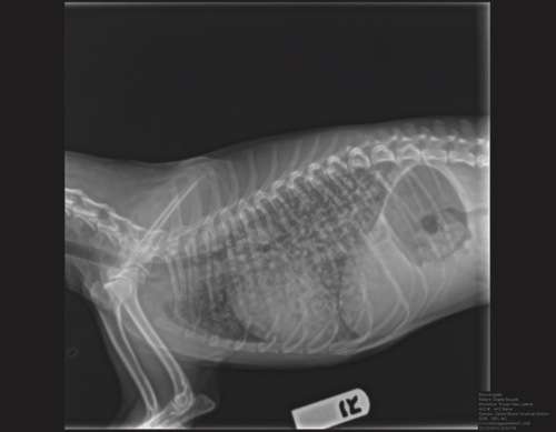
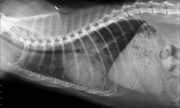
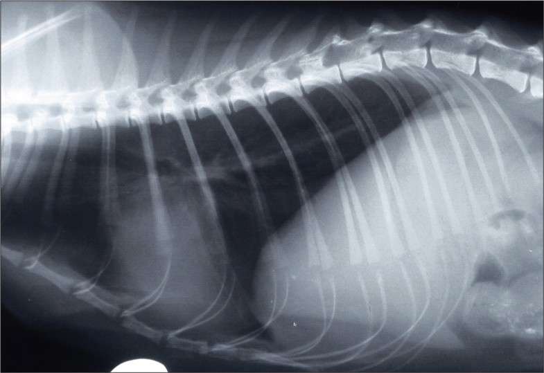
Almost all cats had thoracic radiographs taken during hospitalization. Most cats with radiographically evident pleural effusion had bilateral effusion . Thoracic abnormalities seen were categorized as involving the lung , cardiomegaly , diaphragmatic hernia , thoracic mass , and pneumothorax . Five cats without radiographic pleural effusion were later confirmed to have pleural effusion via thoracic ultrasound. Of the cats that received thoracic ultrasound, most exhibited bilateral pleural effusion . Thirty-two cats that received thoracic ultrasonography were found to have thoracic masses.
Pleural Space Infection Can Become Permanent
Castiel after chest tap, top to bottom X-Ray
The infection not only pockets not the connective tissue lining, not only infects the tissues themselves, but also coats the outside of the lungs and the lining of the chest wall. Also, possibly, whatever injected the infection into the pleural space could still be in the body causing a nidus of infection, or a constant source of new infection. Eve with open chest surgery, placement of a chest drain tube to take out ongoing pus accumulations, and infusion of antibiotics into the pleural space, some cats so not survive the surgery, and some cats do not clear the infection. Or they clear the infection, but there is significant scar tissue left in the pleural space that causes pulling and restriction on the movement and expansion of the lungs that compromises breathing anyway, even if the fluid is all gone.
Dont Miss: Blue Wet Cat Food Review
You May Like: Pellet Sifting Litter Box
Working Up Pleural Effusions In Cats
Cats presenting with pleural effusion are nearly always in respiratory distress, ranging from an increased respiratory rate and effort to open mouth breathing. In the latter situations, therapeutic intervention must be initiated quickly to prevent respiratory arrest. The therapeutic intervention also provides your first diagnostic test.
Cats presenting with pleural effusion are nearly always in respiratory distress, ranging from an increased respiratory rate and effort to open mouth breathing. In the latter situations, therapeutic intervention must be initiated quickly to prevent respiratory arrest. The therapeutic intervention also provides your first diagnostic test. However, the other 2 rule-outs for acute onset of severe respiratory distress is pulmonary edema secondary to cardiac failure and feline asthma. A rapid decision must be made to determine which therapy is most appropriate for the underlying cause.
Diagnosis Of Pleural Effusion In Cats

Diagnostic tests are needed to exclude other diseases that may cause similar signs and to determine the underlying cause of the effusion. Diagnostic tests that your veterinarian may wish to perform include:
You May Like: How To Count Cat Years
What Is The Treatment
Measures to improve the cats respiratory capacity are usually the first steps in therapy. Fluid can be drained from the pleural space with a syringe and needle. In almost all cases, the fluid will reform and a drain tube will need to be surgically implanted to facilitate daily drainage. This tube may be left in place until chyle accumulation stops. If fluid is still accumulating after 1-2 weeks, thoracotomy may be recommended to search for the underlying cause or to repair a torn thoracic duct which will not heal. When the cause is trauma, most cats will heal on their own and surgery will not be needed.
Feline chylothorax is a disease under active research. Some new treatment modalities may be on the horizon. Regardless, the most successful therapy will be directed toward treating the underlying disorder.
Whats The Prognosis For Mediastinal Lymphoma In Cats
Typically, this period of remission lasts only 2-9 months, and then cats become ill again. Mediastinal lymphoma in cats with feline leukemia carries a poor prognosis, with an average survival time of 3 months. In cats without feline leukemia, mediastinal lymphoma often shows at least a partial response to chemotherapy.
Read Also: 17 Cat Years In Human Years
About Dr Joanna Woodnutt Mrcvs
Dr. Joanna Woodnutt is a small animal veterinarian and writer who is passionate about helping owners to learn more about their pets in order to improve animal welfare. She loves to write and wants to empower owners to make the best decisions for their pets by giving them all the information they need. In her spare time, she takes consultations on the small island of Guernsey.
What Is The Treatment For Pleural Effusion
Pleural effusion is a medical emergency and should be treated aggressively as soon as clinical signs occur. Cats have limited ability to handle any condition that compromises their breathing and often deteriorate or decompensate quickly.
The first objective of treatment is removal of the fluid compressing the lungs to allow for more normal lung expansion. This is accomplished by thoracocentesis, which is both a diagnostic procedure and a treatment. The cat often requires oxygen therapy for stabilization prior to performing thoracocentesis. Long-term management is based on the specific cause of the fluid accumulation but includes periodic thoracocentesis, surgery such as thoracic duct ligation, and feeding special low-fat diets or supplements.
Also Check: 3 Month Old Kitten Food
Can A Feline Have Pleural Effusion With Lymphoma
Mediastinal lymphoma is the most common form of lymphoma to be linked to the Feline Leukemia Virus , making that virus a major factor for the eventual development of pleural effusion. If your veterinarian determines there is an effusion, either with scans or by listening to the chest region, he may try to drain the fluid.
Recent Posts
You May Like: How To Get Matts Out Of Cat Hair
Lung Cancer In Cats Treatment
Unfortunately, the prognosis for lung cancer is often poor, meaning that treatment may be palliative rather than focused on curing the disease.
Unfortunately, lung cancer in cats carries a poor prognosis, and treatment is often palliative- to reduce symptoms, rather than cure the disease. Your veterinarian will discuss the treatment options with a veterinary oncologist to determine the best course of action for your cat.
Surgical removal of the primary lung tumour or lung lobe is possible, especially if no metastases have been detected. This involves a thoracotomy- major open-chest surgery that is usually only offered at specialist centers due to the high risks involved. If metastases are present, surgery like this is unlikely to bring enough benefit to make the high-risk procedure worthwhile.
Chemotherapy and radiotherapy is sometimes recommended, especially where metastatic spread has already happened.
If the tumour has caused a pleural effusion, this can be drained to provide some relief.
Read Also: How To Get Matts Out Of Cat Hair
Read Also: Calico Cat Facts
What Is The Prognosis For A Cat Diagnosed With Pleural Effusion
The prognosis will be based on the specific diagnosis and response to initial treatment. A guarded prognosis is always given when initially confronted with a cat experiencing open-mouth breathing. Most cats with chylothorax have a good but guarded long-term prognosis provided the owner complies with the veterinarian’s instructions.
Mediastinal Lymphoma And Pleural Effusion

Lymphoma is a broad category of cancers that can occur in many places within a cat’s body. One such location is the mediastinal area between or in front of the lungs. When lymphoma occurs in this area, it is much more likely to cause a dangerous fluid buildup simply because of its proximity to the lungs and heart. Mediastinal lymphoma is the most common form of lymphoma to be linked to the Feline Leukemia Virus , making that virus a major factor for the eventual development of pleural effusion.
Also Check: Are Gerbera Daisies Poisonous To Cats
Presenting Complaint And History
Presenting complaints included anorexia or decreased appetite , respiratory distress , lethargy , cough , tachypnea , vomiting , gagging , weight loss , and collapse . Duration of clinical signs was both acute and chronic ranging from < 12 h to up to 3 mo. Thirteen animals also had a documented fever , at the time of presentation. Eleven animals had received antimicrobials before confirmation of LLT. Three animals sustained vehicular trauma at 2, 3, or 5 wk before presentation and all 3 had a diaphragmatic hernia and pleural effusion identified on pre-operative imaging or at the time of thoracotomy in addition to the LLT. Cats and dogs appeared to display similar clinical signs. All cats and all but 3 dogs were presented with complaints of respiratory compromise including tachypnea, respiratory distress, and cough. The 3 dogs without respiratory signs had a combination of anorexia, lethargy, and fever and duration of clinical signs ranging from 3 to 10 d.
What Is Pleural Effusion
Pleural effusion refers to the abnormal accumulation of fluid within the chest cavity. In pleural effusion, the fluid is not found within the lungs, but instead within the pleural sac. In pleural effusion, the lungs are floating in a chest that is full of fluid. This fluid occupies space within the chest, keeping the lungs from expanding as fully as they should.
You May Like: What Was The Name Of Hp Lovecrafts Cat
Pleural Effusion In Cats
Pleural effusion is the abnormal accumulation of fluid within the chest cavity, which is lined by a membrane — the pleural lining. This occurs in cats either because too little fluid is being absorbed in the pleural cavity, or because too much fluid is being produced in the pleural cavity. Alterations in the cat’s blood pressure and protein content in the blood, or the penetrability of blood vessels and lymphatic function, may contribute to fluid accumulation.
The condition or disease described in this medical article can affect both dogs and cats. If you would like to learn more about how this disease affects dogs please visit this page in the PetMD health library.
Diagnostic Imaging: Thoracic Radiographs
All 39 animals had thoracic radiographs taken within 72 h of initial presentation to Tufts University or their primary care veterinarian. Twenty-six animals had radiographs taken and interpreted by the diagnostic imaging service at Tufts University, while 13 animals had radiographs taken at their primary care veterinarian before referral. For the 26 animals with radiography done at Tufts University, the most commonly documented radiographic abnormalities included pulmonary consolidation , pleural effusion , vesicular pattern of an individual lung lobe , and lack of visualization of the bronchus or vasculature within the affected lung lobe .2). Of these 26 animals, 15 had LLT as the primary differential diagnosis and 7 were considered to have LLT as a differential diagnosis. In the other 4 animals, LTT was not suspected based on radiographs, although 3 had pleural effusion obscuring visualization of the lung lobes and 1 had a large volume pneumothorax. Interestingly, in only 2 of the 4 cats was LLT suspected based on radiographs alone. Thirteen animals had thoracic radiographs taken at their primary care veterinarian with 8 of these cases suspected of having an LLT either by the referring veterinarian, admitting clinician at Tufts University, or a Tufts University radiologist. Overall, of 39 animals with thoracic radiography, LTT was suspected in 30 cases.
Also Check: Hp Lovecraft Car
What Are The Clinical Signs Of Pericardial Effusion
Signs of pericardial effusion can vary dramatically, depending on the severity and time course of the condition.
Early signs often include abdominal fluid accumulation and the resulting visible abdominal enlargement and exercise intolerance. In some cases, fainting may be noted with physical exertion. Cough and decreased appetite may also be observed. If pericardial effusion persists at mild levels over a long period of time, affected cats may also develop muscle wasting.
In severe cases, especially with an acute onset of disease, pericardial effusion may cause sudden collapse and death with no prior signs.
What Is A Pleural Effusion
Pleural effusion refers to an accumulation of fluid within the pleural cavity. This fluid is usually located within the pleural sac which is a serous membrane covering the lungs. The pleural sac can be thought of as a “plastic wrap” around the lungs that can potentially hold fluid or air . It normally contains only a small amount of fluid, sufficient to keep the lung surfaces slippery and prevent them from sticking to the chest wall.
“…pleural effusion involves fluid on the outside or surrounding the lungs…”
When excess fluid enters this space, it compresses the lungs so that they cannot fully expand and results in impaired breathing. It must be emphasized that a pleural effusion involves fluid on the outside or surrounding the lungs, not within the lung tissues. Fluid accumulation within the lungs is generally called pulmonary edema.
You May Like: What Is The Difference Between Cat Years And Human Years
What Are The Clinical Signs
The main clinical sign of chylothorax is labored breathing. However, this finding is common to all cases of pleural effusion. Some cats appear to be holding their breath because there is a delay between inspiration and expiration. Interestingly, coughing is the first sign of chylothorax in some cats it is not typically found with other causes of pleural effusion. This can be important because there are relatively few causes of coughing in the cat as compared to the dog.
In some situations, clinical signs of the underlying disease may overshadow those of the pleural effusion. Occasionally, owners note no abnormalities other than depression or exercise intolerance. This is because fluid gradually accumulates in the pleural space, and the cat is able to adapt for awhile.
How Long Can Pleural Effusion Last

Most people recover within a few days or weeks. Minor complications from more invasive treatments can include slight pain and discomfort, which often go away with time. Some cases of pleural effusion can have more serious complications, depending on the severity of the condition, cause, and treatment used.
Don’t Miss: How Often Should An 8 Week Old Kitten Eat
How Is The Condition Diagnosed
Diagnosis is based on medical history and clinical signs, the presence of fluid in the chest cavity on thoracocentesis , chest radiographs and chest ultrasound. Blood and urine tests are often performed to look for any underlying infectious or systemic disease that is causing the fluid accumulation. The fluid removed from the chest cavity is analyzed for clues as to the origin or cause.
How Is The Diagnosis Made
The following tests are usually performed to achieve a diagnosis of chylothorax.
Once the presence of chylothorax is confirmed, additional tests will be performed in an attempt to identify an underlying cause. Such tests include routine blood work, additional chest and abdominal radiographs, tests for leukemia virus and immunodeficiency virus, cardiac tests, and heartworm testing.
Also Check: What Is A Cat Year Equal To In Human Years
What Is Fluid In The Chest
Fluid in the chest is also known as pleural effusion. This occurs when fluid is present outside the lungs, in the space between the lungs and the chest wall. Normally, this area only has a small amount of fluid simply to keep the lungs from adhering to the chest wall. When excessive amounts of fluid accumulate, serious complications can arise because the cats lungs cannot expand properly. This is a potentially life-threatening situation for your cat and emergency treatment is necessary.
While most cats are generally healthy, some can develop conditions that can compromise their health and well-being. Cats can develop fluid in the chest as a result of various conditions. Regardless of the cause, fluid in the chest in cats can be very serious.
Vet bills can sneak up on you.
Plan ahead. Get the pawfect insurance plan for your pup.
Fluid In The Chest Average Cost
From 565 quotes ranging from $500 $4,000
Average Cost
Dont Miss: What Is A Cats Normal Heart Rate
Is Pleural Effusion Serious
The seriousness of the condition depends on the primary cause of pleural effusion, whether breathing is affected, and whether it can be treated effectively. Causes of pleural effusion that can be effectively treated or controlled include an infection due to a virus, pneumonia or heart failure. Two factors that must be considered are treatment for associated mechanical problems as well as treatment of the underlying cause of the pleural effusion.
Dont Miss: Is Blue Wilderness A Good Cat Food
Also Check: How Do Cats Age Vs Humans