When Ct Scans Are Used
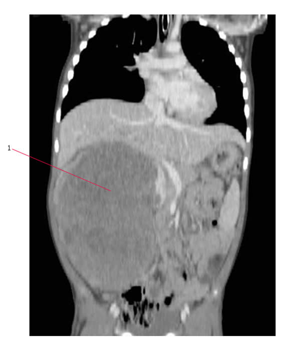
CT scans can produce detailed images of many structures inside the body, including the internal organs, blood vessels and bones.
They can be used to:
- diagnose conditions including damage to bones, injuries to internal organs, problems with blood flow, stroke, and cancer
- guide further tests or treatments for example, CT scans can help determine the location, size and shape of a tumour before having radiotherapy, or allow a doctor to take a needle biopsy or drain an abscess
- monitor conditions including checking the size of tumours during and after cancer treatment
CT scans wouldn’t normally be used to check for problems if you don’t have any symptoms .
This is because the benefits of screening may not outweigh the risks, particularly if it leads to unnecessary testing and anxiety.
What Does The Ct Equipment Look Like
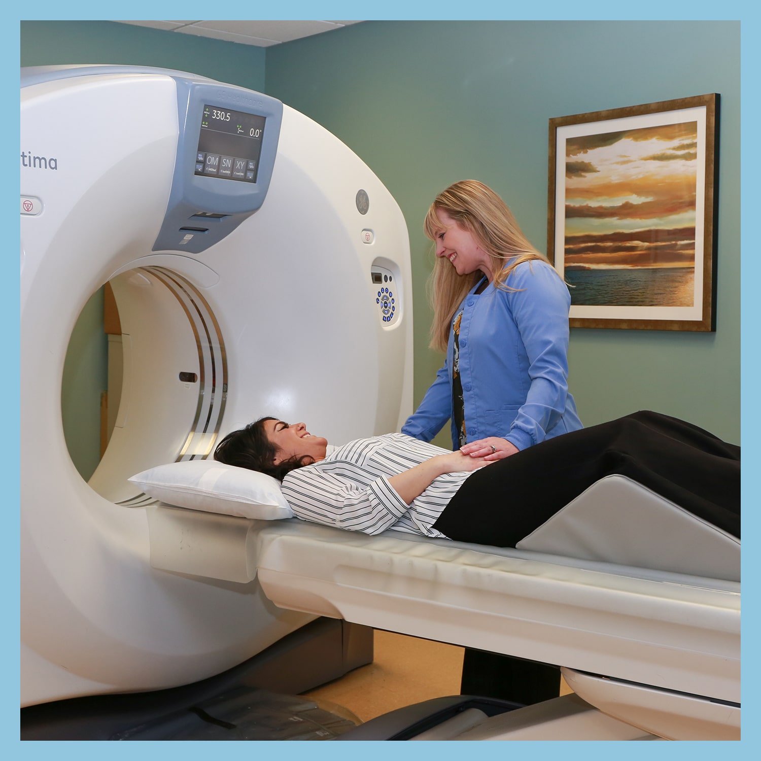
The CT scanner is typically a large, donut-shaped machine with a short tunnel in the center. You will lie on a narrow table that slides in and out of this short tunnel. Rotating around you, the x-ray tube and electronic x-ray detectors are located opposite each other in a ring, called a gantry. The computer workstation that processes the imaging information is in a separate control room. This is where the technologist operates the scanner and monitors your exam in direct visual contact. The technologist will be able to hear and talk to you using a speaker and microphone.
How Does Ct Work
Unlike a conventional x-raywhich uses a fixed x-ray tubea CT scanner uses a motorized x-ray source that rotates around the circular opening of a donut-shaped structure called a gantry. During a CT scan, the patient lies on a bed that slowly moves through the gantry while the x-ray tube rotates around the patient, shooting narrow beams of x-rays through the body. Instead of film, CT scanners use special digital x-ray detectors, which are located directly opposite the x-ray source. As the x-rays leave the patient, they are picked up by the detectors and transmitted to a computer.
Each time the x-ray source completes one full rotation, the CT computer uses sophisticated mathematical techniques to construct a two-dimensional image slice of the patient. The thickness of the tissue represented in each image slice can vary depending on the CT machine used, but usually ranges from 1-10 millimeters. When a full slice is completed, the image is stored and the motorized bed is moved forward incrementally into the gantry. The x-ray scanning process is then repeated to produce another image slice. This process continues until the desired number of slices is collected.
Don’t Miss: Best Places To Cater From
What Are Examples Of Nibib
Imaging for acute ischemic stroke: Stroke, which can have lasting neurological injuries, is also a leading cause of death worldwide. To mitigate damage to the brain, patients may receive endovascular treatment, where the clot blocking the blood supply is either removed or dissolved. However, identifying patients who will benefit from endovascular therapy, such as those with only a small volume of irreversibly injured brain tissue, remains challenging, and time is a critically important factor for a successful clinical outcome.
NIBIB-funded researchers have developed an image reconstruction technique to more efficiently triage patients who present with symptoms of a stroke. This CT-based method can be used to rule out the presence of a hemorrhage to find the site of the blood clot and to identify the extent of damaged brain tissue. Such a technique could significantly shorten the time from the diagnosis of a stroke to the start of endovascular therapy, and could also guide the endovascular treatment. Following evaluation in animal models, researchers plan to validate this CT imaging technique in human studies.
For more information about CT, watch our video here.
Large Bore Ct Scanner/radiation Therapy With Simulation

This health-care tool produces detailed images of areas inside the body for planning radiation therapy. It uses simulation, fluoroscopy and respiratory gating to plan and deliver radiation.
Advantages may include:
- Locating abnormalities and guiding precision radiation therapy
- Planning for treatment options based on a patients breathing patterns
- Allowing for varying patient sizes and positioning
- Spending 15 to 30 minutes for the session, including the time it takes to put you in position and set up the equipment
Don’t Miss: Cat Skid Steer Quick Attach Parts
Benefits Of A Ct Scan
- CT scans have shown to be a cost-effective imaging tool for a wide range of clinical problems.
- CT scanning is painless, noninvasive and accurate.
- New state-of-the art CT scanners produce superior exams using a fraction of time and radiation exposure. At HSS, we practice ALARA protocols for all our patients, including in children. New technologies make faster scanning possible. For children this means shorter imaging times and less time required to hold still in order to produce clear, crisp images. Also, shorter scan times will make it easier for children and adults to hold their breath during critical parts of the exam. Scanning helps to reduce the need for sedation in children.
- A major advantage of CT is its ability to image bone, soft tissue and blood vessels all at the same time.
- Compared to , CT is less sensitive to patient movement and can be performed if you have an implanted medical device of any kind.
- CT imaging provides real-time imaging, making it a good tool for guiding minimally invasive procedures such as needle biopsies and needle aspirations. Sometimes is substituted for CT as a method of image guidance for procedures, especially in children.
- A diagnosis determined by CT scanning may eliminate the need for exploratory surgery and surgical biopsy.
Reporting Problems To Fda
Prompt reporting of adverse events can help the FDA identify and better understand the risks associated with the product. We encourage health care providers and patients who suspect a problem with a medical imaging device to file a voluntary report through MedWatch: The FDA Safety Information and Adverse Event Reporting Program.
Health care personnel employed by facilities that are subject to Reporting Adverse Events should follow the reporting procedures established by their facilities.
Medical device manufacturers, distributors, importers, and device user facilities must comply with the Reporting Adverse Events .
In addition to following the general recommendations for reporting problems for adverse events associated with CT overexposure, the following information should be included in reports, if available:
- the protocol you were following during the event
- the CT conditions of operation and
- the dose-index values displayed .
Don’t Miss: Do Cats Help With Depression
What Should I Do To Prepare For The Examination
If your CT scan will be performed as part of another procedure , follow the doctor’s instructions for that procedure. If the CT scan is to include an injection of a contrast agent, it is advisable that you do not eat or drink for a several hours before the procedure. If you have any allergies, especially allergies to medications, contrast agent, local anesthesia, Betadine soap, or latex, be sure to inform your physician at the time of scheduling of the procedure, and also inform the CT technologist and the radiologist before the start of the procedure.
If you are pregnant or think you may be pregnant, be sure to inform your physician, the technologist, and the radiologist prior to the procedure. Most procedures/examinations using X-rays will not be performed on pregnant women unless the benefits of the procedure/examination outweigh the risks of radiation exposure to the fetus.
What Are The Limitations Of Body Ct Scanning
Soft-tissue details in areas such as the brain, gallbladder, internal pelvic organs, and joints can often be better evaluated with magnetic resonance imaging . In pregnant women, while CT can be performed safely, other imaging exams not involving radiation, such as ultrasound or MRI, are preferred but only if they are likely to be as good as CT in diagnosing your condition.
A person who is very large may not fit into the opening of a conventional CT scanner. Or, they may be over the weight limitusually 450 poundsfor the moving table.
Read Also: Blue Buffalo Wilderness Chicken Cat Food
What Can I Expect After Getting A Ct Scan
If intravenous contrast dye was used, you may have soreness at the site of IV line for up to a few hours. If a biopsy, joint injection, and/or aspiration, or nerve root block was performed, soreness at the site of needle placement may last up to a few days or a hematoma with swelling and a black and blue appearance can develop at the site of needle placement. If you had a therapeutic injection you will receive a discharge instruction form.
What Will I Experience During And After The Procedure
CT exams are generally painless, fast, and easy. Multidetector CT reduces the amount of time that the patient needs to lie still.
Though the scan is painless, you may have some discomfort from remaining still for several minutes or from placement of an IV. If you have a hard time staying still, are very nervous, anxious, or in pain, you may find a CT exam stressful. The technologist or nurse, under the direction of a doctor, may offer you some medication to help you tolerate the CT exam.
If the exam uses iodinated contrast material, your doctor will screen you for chronic or acute kidney disease. The doctor may administer contrast material intravenously , so you will feel a pin prick when the nurse inserts the needle into your vein. You may feel warm or flushed as the contrast is injected. You also may have a metallic taste in your mouth. This will pass. You may feel a need to urinate. However, these are only side effects of the contrast injection, and they subside quickly.
If you swallow oral contrast material, you may find the taste mildly unpleasant. However, most patients can easily tolerate it. If you receive an enema, you can expect to experience a sense of abdominal fullness. You may also feel an increasing need to expel the liquid. If so, be patient the mild discomfort will not last long.
With pediatric patients, a parent may be allowed in the room but may need to wear a lead apron to minimize radiation exposure.
Read Also: Cat Puking And Not Eating
Are Crumpets High In Fat
A 100-gram serving of crumpets generally contains between 1 and 2 grams of fat, though crumpets with added ingredients may contain more. When choosing a crumpet, remember that the American Heart Association recommends that you limit your intake of fat to 25 to 35 percent of your total caloric intake.
How Does A Pet Scan Work

During a PET scan procedure, a technologist will inject a small amount of the radioactive tracer into a patients vein, usually on the inside of an elbow. After the injection, the tracer will move through the body and gather in tissues and organs. A patient will need to wait for about an hour for their body to absorb the tracer. Tracers will collect in tumors, inflamed areas or cancer cells for doctors to see.
After the tracer is absorbed, the patient will move to an exam table that slides into a scanner. The PET scanner will detect the tracer and turn the data into 3-D images on a computer monitor. Some imaging centers combine PET scans with CT scans to create specialized images.
Read Also: Bengal Cat Rescue New England
How Are Pet And Ct Scans Similar
CT scans and PET scans focus on different aspects of the human body and use unique materials to do so, but they still share a lot in common. Similarities between CT scans and PET scans include:
- Both are usually performed at an outpatient facility and do not require a hospital stay.
- Both can be used to detect cancer.
- Both are accurate, painless and noninvasive procedures.
- Both help eliminate the need for exploratory surgery.
Most importantly, both types of imaging tests help doctors diagnose the cause of a patients symptoms. This allows doctors to effectively treat their patients and help them enjoy healthy lives.
What Is The Difference Between A Ct And Pet Scan
Perhaps the main difference between a CT scan and a PET scan is their focus. A CT scan creates a detailed non-moving image of organs, bones and tissues. A PET scan, on the other hand, shows doctors how the tissues in your body work on a cellular level. Other differences include:
- They use different materials: CT scans pass x-rays through the body to create images. A PET scan uses a radioactive material which emits energy. The energy is then detected by a special camera to produce images.
- A PET scan is more time-consuming: A CT scan is performed in minutes. This makes it an excellent tool in emergency situations when doctors need to act fast. A PET scan could take 20 minutes to several hours, depending on a patients condition. In some cases, the procedure may be performed over several days.
- Radiation does not stay in the body with a CT scan: After a CT scan, no radiation remains in the body. On the contrary, a small amount of radiation may stay in the body for a short time after a PET scan.
- PET scans detect cancer earlier than other tests: Unlike other forms of imaging, a PET scan shows molecular activity and helps doctors identify diseases in the earliest stages. For this reason, a PET scan is a highly reliable tool for detecting cancer. CT scans show signs of an issue after a disease begins to change the structure of tissues or organs.
Also Check: Best Dry Cat Food For Hairballs
Information For Health Care Providers
The FDA has regulations covering the safety and effectiveness and radiation control of all X-ray imaging devices, including CT. Individual states and other federal agencies regulate the use of CT devices through recommendations and requirements for personnel qualifications, quality assurance and quality control programs, and facility accreditation.
Ct Scan With Or Without Contrast
Some types of CT scans require the use of a special chemical called contrast, which helps make soft tissue easier to see on the images. Although you might have heard contrast called a dye, it doesnt change the color of the organs or soft tissues inside the body. Instead, it works by blocking the x-rays. Contrast looks white on the images, making the organs or other tissues stand out.
Not all types of CT scans require contrast. Your doctor will let you know if yours does before you schedule it. Since some people are allergic to the ingredients commonly used in contrast, such as iodine or barium, its crucial to tell your doctor about any known allergies before they schedule the scan.
There are different ways of receiving contrast before a scan. You might get it intravenously, through an injection. Your technologist might also give you something to swallow. If you are going to have contrast before a CT scan, you wont be able to eat or drink anything starting several hours beforehand.
Recommended Reading: Can Dogs Be Allergic To Cats
Pet Scan Vs Ct Scan: Use In Cancer Imaging And Treatment
Oncologists may order both PET scans and CT scans for their cancer patients. Both scans can help monitor the impact of cancer treatment on the body. The PET scan shows the presence of cancer cells throughout the body but a CT scan will reveal abnormal tissue and detail images, says Cleveland Clinic. It is now common for cancer patients to visit PET/CT imaging centers. This allows the opportunity for patients to get both tests at the same time.
Ct Scans Of The Tummy
If you are having a CT scan of your abdomen, you might need to:
- drink a liquid contrast medium some time before the scan
- drink more of the liquid contrast or water in the x-ray department
- stop eating or drinking after midnight the night before the scan
You usually have the contrast medium by injection and also as a drink. This helps to show up the gut more clearly in the scan.
You May Like: Cat Food High In Fiber
Schedule Your Appointment Today
Whether your doctor has ordered a CT scan or a PET scan, you may feel a little anxious about the procedure. Any medical imaging exam can cause worrisome feelings before and after the test. At Health Images, our team of dedicated, compassionate technologists want to make the experience as pleasant and stress-free as possible.
We have the latest technology and technologists who care, so you can depend on us to meet your imaging needs. To learn more about our services or schedule an appointment, contact a Health Images center today.
Which Type Of Scan Is Best For You

CT scans, also known as computed tomography scans, are considered to be the gold standard for imaging because they provide a detailed picture of the inside of the body. CAT scans, on the other hand, are more widely used because they can be completed in less time and are less expensive. Heres a closer look at the two types of scans:
CT Scan: CT scans use a series of X-rays to create an image of your body. They can penetrate deeply into your tissues, so theyre best used for images that need detail, such as tumors or injuries. CT scans are also good for checking for fluid buildup in the brain and other areas of the body.
CAT Scan: CAT scans use a beam of radiation to create an image. The radiation is directed at objects close to the scanner, which means that it cant penetrate as far as CT scans. That means CAT scans are good for images that need wide coverage, like whole body images or images of organs.
You May Like: Cat Food Pate Vs Chunks
What Is A Ct Scan For
A CT scan produces images using an X-ray tube that travels 360 degrees around your body. As it moves, it gathers information from multiple angles to create cross-sectional images.
Doctors order CT scans for a number of reasons, including to:
- Look for bone and joint problems such as complex fractures and tumors
- Detect masses and spots associated with conditions like heart disease, cancer and emphysema
- Locate clots, tumors, infection or fluid buildup
- Check for internal injuries and bleeding following a traumatic event like a car accident
- Provide information in preparation for a surgery, biopsy or other procedure
- Compare images taken over a period of time to gauge the effectiveness of a treatment such as the chemotherapy or radiation used to shrink a tumor
In some cases, whats called a CT scan with contrast is needed. With this procedure, the technician performing the procedure inserts a catheter into one of your arms in order to administer a special dye called contrast material. This dye makes it easier for the scan to create a crisp image of tissues and organs that are less dense than bone and therefore may not show up as clearly.
The detail in a CT scan with contrast may make it the preferred approach over other imaging techniques in some instances. For example, a CT scan can provide better information on the bones of the spine than either a standard X-ray or an MRI scan, which is helpful when assessing conditions that affect the vertebrae and other bones.